Science Class 9 Notes From Chapter 6 – Tissues
Q 1: Define tissue. Write names of types of tissues in humans.
A 1: A group of cells that are similar in structure and/or work together to achieve a particular function forms a tissue.
Following are the four types of tissues found in human body:
- Epithelial tissue.
- Connective tissue.
- Muscular tissue.
- Nervous tissue.
Q 2: What is a unicellular organism? Give examples of unicellular organisms.
A 2: Single celled organisms are called unicellular organisms. Remember "Uni" means single. Some examples of unicellular organisms are amoeba, paramecium, bacteria, fungus and plasmodium.
Q 3: What is a multicellular organism? Give two examples.
A 3: Multi means many, so multicellular organisms have multiple cells.
Examples of multicellular organisms are plants, animals, humans etc.
Q 4: What is the function of muscle cells in humans?
A 4: The function of muscle cells in humans is to cause movement and produce force.
Q 5: What is the function of nerve cells in humans?
A 5: The function of nerve cells is to carry messages from various body parts to brain and back by electrical impulses. Nerve cell is also known as neuron.
Q 6: Which tissues in plants conduct food and water from one part or the plant to other parts?
A 6: Xylem vessels conduct water and mineral while phloem tubes conduct water in a plant.
Q 7: Are plants and animals made of same type of tissues?
A 7: No, plant and animals are made of different types of tissues.
Plants are made of following types of tissues:
Meristematic
Permanent
Protective
Complex
Animal tissues are made of:
Epithelial
Muscular
Nervous
Connective
Q 8: Write three differences in plants and animal tissues?
A 8:
S. No.
|
Plants Tissue
|
Animals tissue
|
1.
|
Most of the tissues are supportive and dead
|
1. Most of the tissues are living.
|
2.
|
Cells need less maintenance
|
2. Cells need more maintenance
|
3.
|
There is limited growth in certain regions
|
3. There is growth in all regions.
|
Q 9: What is a meristematic tissue? Write their types and function in brief.
A 9: Meristematic tissues or simply called meristems are tissues in which the cells remain forever young and divide actively throughout the whole life of the plant.
When a meristematic cell divides in two, the new cell that remains in the meristem is called an initial, the other the derivative.
Meristematic cells are generally small in size and cuboidal shaped with large nuclei, small vacuoles, and thin walls.
They are of three types:
Apical meristem: They are present at the tips of stems, roots, and branches. They are responsible for the axial growth in a plant.
Intercalary meristem: They are present at the base of internodes, and are responsible for the growth of internodal region.
Lateral meristem: They are present on the lateral side of stems and roots. Lateral meristem is responsible for the radial growth of plants. Vascular cambium and cork cambium are examples of lateral meristem.
Q 10: With help of a diagram show the location of meristematic tissue in plant body.

A 10: The diagram on left shows the location of meristematic tissue in plant body. It shows Apical meristem, Intercalary meristem and Lateral meristem.
Q 11: Write functions of different types of meristematic tissues in brief.
A 11: Apical meristem - Meristem at the tip of a plant shoot or root causes the shoot or root to increase in length. It is responsible for primary growth of plant.
Intercalary Meristem: It is the meristem that occurs between permanent tissues. It represents the remains of the apical meristem. It is particularly common at the nodal regions. It may also occur at the base of the leaves. The intercalary meristem also contributes towards the increase in length as it brings about elongation of the internodal regions. It is also responsible for the formation of branches at the nodal regions.
The apical and intercalary meristems are examples of primary meristem.
Lateral Meristem: It appears in the mature tissues of roots and shoots. It is also called the secondary meristem as it appears later in a plant’s life. It helps in adding secondary tissues to the plant body and in increasing the thickness of stem of plants. Lateral meristem tissues are very active as they have dense cytoplasm, thin cellulose walls and prominent nuclei.
Q 12: What is the function of vacuoles?
A 12: A vacuole is a temporary stoage area in a cell. Vacuoles are membrane-bounded compartments within some eukaryotic cells that can serve a variety of secretory, excretory, and storage functions.
In general, vacuole functions include:
In general, vacuole functions include:
- Removing unwanted structural junk material
- Isolating materials that might be harmful to the cell
- Containment of waste products
- Maintaining internal hydrostatic pressure or tension within the cell
- Maintaining an acidic internal pH (measure of acidity or basicity)
- Containing small molecules
- Exporting unwanted substances from the cell
- Enabling the cell to change shape
- Aids in destruction of invading bacteria or of misfolded proteins that have begun to build up within the cell.
Q 13: Write feaures of meristematic tissue.
A 13: The cells of the meristematic tis sues are quite distinct in their formation, structure and functional characteristics from other cells. The following are the characteristics of the meristematic cells:
- Cells have the power of active division
- They are compactly arranged in tissue and there is absolutely no intercellular space
- The cell wall is thin and primary in nature, containing only cellu lose. It is uniformly thick. There is no secondary thickening
- Cells possess dense protoplasm with a prominent large nucleus compared to other cells of equal volume
- Vacuoles are small or absent to tally
- Mitochondria and endoplasmic reticulum are very little differentiated
- Cells contain relatively more number of ribosomes
- Cells are metabolically very active
Q 14: Define permanent tissue. Give names of some of permanent tissues in plants.
A 14: Permanent tissue is the tissue which makes the plant hard and stiff. Permanent tissues take up a specific role and lose the abiltiy to divide.
Some of permanent tissues are simple and complex.
Q 15: Define differentiation.
A 15: The process of taking up a permanent shape, size and a function is called differentiation.
Q 16: Write in brief about parenchyma, chlorenchyma, aerenchyma, collenchyma and sclerenchyma.
 A 16: Parenchyma - A permanent tissue of few layer of cells that forms the basic packing tissue.
- Consists of relatively unspecialised cells with thin cell walls
- Live cells
- Usually loosely packed so that large spaces are there
- Provides support to plants and also stores food.
- Parenchyma of stems and roots also stores nutrients and water.
 Chlorenchyma
In some situations simple permanent tissue contains chlorophyll and performs photosynthesis then it is called chlorenchyma.

Aerenchyma
Is a parenchyma type of tissue found in aquatic plants with large cavities that give buoyancy to the plants to help them float.
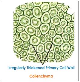
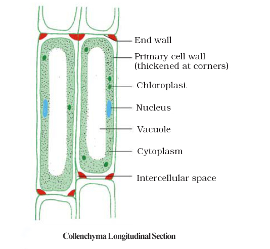
Collenchyma
- A permanent tissue that gives flexibilty to plants.
- Allows easy bending in various aprts of a plant – leaf and stem, without breaking.
- Provides mechanical support to plants.
- Found in leaf stalks (stem or main axis) below the epidermis (outermost layer)
- Living elongated and irregularly thickened at the corners with very less intercelluler space.
Sclerenchyma

 A 31: Both Xylem and Phloem are conducting tissues and part of transport system that carry fluids (liquid and gas) in plants (vascular bundles). They are complex permanent tissues made of more than one type of cells.

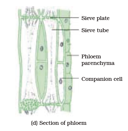
Q 33: Define sieve tubes.
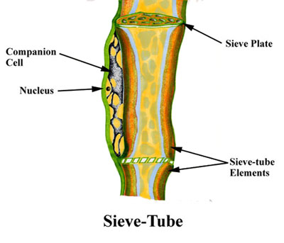 A 33: Sieve tubes are element of phloem tissue consisting of a longitudinal row of thin-walled elongated cells with holes in their connecting walls through which food materials passes
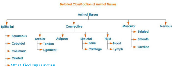
Q 34: What are different kinds of animal tissues? Write their names and in brief about them.

- Type of permanent tissue that makes the plant hard and stiff
- Husk of coconut is made of sclerenchymatous tissue
- Cells of this tissue are dead
- They are long, narrow as there walls are thickened due to lignin
- Present in stems, around vascular bundles, in the veins of leaves and in the hard covering of seeds and nuts
- Provides strength to the plant parts

Q 17: Define lumen in biology reference
A 17: The inside space in a tubular structure is called lumen.
Q 18: Define lignin.
A 18: A chemical substance which acts as cement and hardens tissue is called lignin.
Q 19: What are guard cells and epidermal cells? Draw their diagram.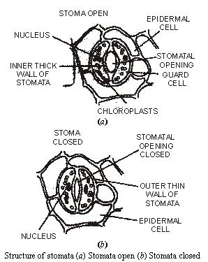
A 19: Guard cells are one of the paired cells in the epidermis of a plant that control the opening and closing of a stoma of a leaf.
Q 20: Write in brief about epidermal cells.
A 20: Epidermal cells are cells on the outermost layer of a plant tissue.
- Epidermal cells on the aerial (in air) parts of the plant often secrete a waxy, water resistant layer on their outer surface. This aids in protection against loss of water, mechanical injury and ivasion by parasitic fungi.
- Cells of epidermal tissue form a continuous layer without intercellular spaces.
- Most epidermal cells are relatively flat.
- Often their outer and side walls are thicker than the inner wall.
- Epidermal cells of the roots, whose function is water absorption, commonly have long hair like parts that greatly increase the total absorptive surface area.
Q 21: What are stomata? Write it functions.
A 21: Stomata are small pores in the epidermis of the leaf.
Stomata are closed by two kidney shaped cells called guard cells.
The word stomata mean “mouth”. These small pores found in the leaves of the plant helps in gaseous exchange during photosynthesis and respiration. Stomata consist of two types of cells, the stoma or the pore and guard cells. Stomata are guarded pair of crescent shaped specialized parenchyma cells called guard cells which regulates the size of opening or pore of stomata.
The main functions of stomata are:
- Exchange of atmospheric oxygen and carbon dioxide
- Transpiration.
Atmospheric air containing carbon dioxide and oxygen enters the plant via these openings, which is used in photosynthesis and respiration. Oxygen produced by photosynthesis in the leaf exits through these openings.
Q 22: Define transpiration. What is its role in plants?
A 22: Water vapour is released into the atmosphere in plants through stomata by a process called transpiration. This helps in cooling the plant and to enable a constant flow of water to travel through the plant tissues and to carry nutrients and other chemicals to the plant tissues.
Q 23: What is photosynthesis? Which gas is needed for the same?
A 23: 'Photo' means “light” and 'synthesis' means preparation. Thus, photosynthesis is the process by which the green plants (having chlorophyll) use light energy of the sun to synthesize carbohydrates.
Photosynthesis is a process having series of biochemical reactions which can be essentially summarised as follows:

Thus, photosynthesis can be defined as a process which utilises carbon dioxide and water in the presence of sunlight and chlorophyll to synthesize carbohydrates like glucose.
In real equation form, photosynthesis can be written as:
6 CO2 + 12 H2O + light energy --> C6H12O6 + 6 O2 + 6 H2O
Q 24: What is cutin?
A 24: Cutin is waxlike, water-repellent polyester consisting of fatty acids that occurs naturally in the walls of many plant cells.
Cutin acts together with wax to form the cuticle, a barrier protecting the above ground surfaces of plants from water loss and microorganisms attack.
Q 25: Define meristem.
A 25: Meristem is a plant tissue responsible for growth, whose cells divide and differentiate (process of taking permanent shape) to form the tissues and organs of the plant. Meristems occur within the stem and leaves and at the tips of stems and roots
Q 26: Is the outer layer of a branch of a tree different from the outer layer of a young stem? How?
A 26: Yes the outer layer of a tree is different from the outer layer of a young stem.
In trees and old plants, a strip of secondary meristem replaces the epidermis of the stem. Cells on the outside are cut from this layer.
This forms the several layer thick cork or the bark of the tree.
Cells of cork in trees are dead and compactly arranged without intercellular spaces.
Outer layer of a tree or cork have a chemical called suberin in their walls that make them impervous (does not allow to pass through) to gases and water.
Q 27: What is suberin?
A 27: A non reactive impermeable waxy substance present in the cell walls of corky tissues is called suberin.
Q 28: Draw a digram to show protective tissue.
A 28: The diagram of protective tissue with cork cells and ruptured epidermis is shown above.
Q 29: What are simple permanent tissues?
A 29: Simple permanent tissues (shown above) are called simple because they are composed of similar types of cells which have common origin and function.
Examples are Parenchyma, Collenchyma, Sclerenchyma and Epidermis.
Q 30: Define complex tissues. Give examples.
A 30: Complex tissues are another type of permanent tissues that are made of more than one type of cells.
All cells of complex tissues coordinate to perform a common function.
Examples are Xylem and Phloem.
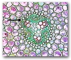
Q 31: What are xylem and phloem? Write differences between them. Make diagrams too.
A 31: Both Xylem and Phloem are conducting tissues and part of transport system that carry fluids (liquid and gas) in plants (vascular bundles). They are complex permanent tissues made of more than one type of cells.
Xylem consists of tracheids, vessels, xylem parenchyma and xylem fibres. Xylem transports water and minerals vertically.
Xylem parenchyma stores food and helps in sideway conduction of water.
- Phloem is a tubular structure conducting tissue in plants that is made up of four types of elements: sieve tubes, companion cells, phloem fibres and the phloem parenchyma.
- Sieve tubes are tubular cells with perforated (having holes) walls.
- In phloem, materials can move in both directions in it.
- Phloem transports food from leaves to other parts of the plant.
Q 32: Define tracheids and vessels. Write one difference between them.
A 32: Tracheids are type of water-conducting, supporting cells having tubular structure in the xylem that lacks perforations in the cell wall.
Q 33: Define sieve tubes.
A 33: Sieve tubes are element of phloem tissue consisting of a longitudinal row of thin-walled elongated cells with holes in their connecting walls through which food materials passesQ 34: What are different kinds of animal tissues? Write their names and in brief about them.
A 34: Different kinds of animal tissues with names and functions in brief are given:
- Epithelial tissue
- Connective tissue
- Muscular tissue
- Nervous tissue
Epithelial tissue
- Covering or protective tissues in the animal body
- Forms a barrier to keep different body systems separate. Examples are skin, the lining of mouth, the lining of blood vessels, lung alveoli and kidney tubules.
- Tightly packed and form a continuous sheet.
- Have a small amount of cementing material between them and almost no intercellular spcaes
- Selectively permeable
Simple Squamous Epithelial cells
- Extremely thin and flat and dorm a delicate lining. Examples are the oesophagus and lining of the mouth and the skin.
- Arranged in many layers to prevent wear and tear
Stratified Squamous Epithelium
- Arranged in a pattern of layers
- Columnar Epithelial Tissue
- Pillar like, facilitates movement across epithelial barrier
Ciliated Columnar Epithelia
- Is columnar epithelia tissue has cilia, i.e hair like projections on the outer surfaces of epithelial cells.
- Cilia can move and their movement pushes the mucus (slippery substance and protective lubricant) forward to clear it.
Connective Tissue
- Connects various parts together
- Cells are loosely spaced and embedded in an intercellular tissue which may be jelly like fluid, dense or rigid. Examples are blood, bones, cartilage and areolar connective tissue
Muscular Tissue
- Consists of elongated cells called muscle fibres
- Responsible for movement in our body
- Contains special proteins called contractile proteins which contract and relax to cause movement
- Types of muscle fibres are striated, smooth and cardiac
Nervous Tissue
- Highly specialised cells for being stimulated and then transmitting the stimulus very rapidly from one place to another
- Examples are brain, spinal cord and nerves
Q 35: What is the function of blood in human body?
A 35: Following are the functions of blood, a type of connective tissue in our body:
- Oxygen is transported to all the body cells through blood.
- Blood flows and carries various substances from one part of the body to the other.
- Food is transported to all cells via blood.
- Blood collects wastes from all parts of the body and carries them to the liver and kidney for disposal.
Q 36: With the help of a chart, show various types of animal tissues.
A 36: Various animal tissues are shown in the given below chart:
Q 37: What is a connective tissue? Write various types of connective tissues.
A 37: A connective tissue is a type of animal tissue that connects various parts of the body. Some types of connective tissues are:
- Areolar tissue
- Adipose tissue
- Compact bone
- Hyaline cartilage
Q 38: Give examples of connective tissues.
A 38: Some examples of connective tissues in animals are blood, bones, cartilage and areolar.
Q 39: Where is areolar connective tissue found?
A 39: Areolar connective tissue is found between the skin and muscles, around blood vessels and nerve.
Q 40: Write types of blood cells in human body.
A 40: The different types of blood cells in human body are:
- Neutrophil (First immune cells)
- Eosinophil (Participate in engulfing and killing bacteria and other microorganisms such as parasites)
- Basophil (Plays role in not allowing the blood to clot too quickly and promotes blood flow to tissues)
- Lymphocyte (To fight off infections by creating antibodies)
- Monocyte (Multiple roles in immune function)
- Platelets (prevent bleeding by clotting)
Q 41: What is the full form of RBC, WBC?
A 41: RBC – Red Blood Cells and WBC – White Blood Cells
Q 42: What is the function of platelets in our body?
A 42: The function of platelets is to clot (a compact group) and protect from bleeding.
Q 43: What do you understand by plasma?
A 43: Blood has fluid tissue called plasma in which RBCs, WBCs and platelets are suspended.
Plasma contains proteins, salts and hormones.
Q 44: What is a ligament? Give example.
A 44: A ligament is a type of connective tissue that is very elastic, has considerable strength and has less tissue.
Q 45: What is a tendon? Give example.
A 45: Tendon means to stretch. It is a type of tissue by which a muscle attaches to bone. A tendon is somewhat flexible, but fibrous and tough.
Q 46: What is the difference between tendon and ligament?
A 46: Tendons are like ligaments in being tough, flexible cords. But tendons differ from ligaments in that tendons extend from muscle to bone whereas ligaments go from bone to bone as at a joint.
As compared to ligament, tendons are fibrous with great strength but limited flexibility.
Q 47: Write in brief about cartilage.
A 47: Cartilage is a kind of soft connective tissue that has widely spaced cells. It is a solid matrix (material between cells) composed or proteins and sugars.
Cartilage smoothens bone surfaces at joints and is also present in the nose, ear, trachea (windpipe) and larynx (voice box).
Q 48: What are fats and where are they stored in human body?
A 48: Fats are large number of oily compounds that are widely found in plant and animal tissues and serve mainly as a reserve source of energy.
In human body, fat, or adipose tissue, is deposited beneath the skin and around the internal organs, where it also protects and insulates against heat loss.
Q 49: Write in brief about muscular tissue.
A 49: Muscular tissue consists of elongated cells also called muscle fibres. Muscular tissue is responsible for movement in our body.
This movement is done as muscles contain special proteins called contracticle proteins which contract and relax to cause movement.
Q 50: Write names of various muscle fibres. Explain about them in brief and show their pictures.
A 50: There are three types of muscle fibres:
- Striated
- Smooth
- Cardiac
Their details in brief are:
Striated
- Shows alternate light and dark bands or striations on staining.
- Are long, cylidrical, unbranched and multinucleate (having many nuclei)

Smooth
- Involuntary muscles, controls movements in the alimentary canal
- Invlountary muscles that control contraction or relaxation of blood vessels

Cardiac
- Involuntary muscles in heart that helps in contraction and relaxation throughout life.
- Are cylindrical, branched and uninucleate.
Q 51: What do you understand by voluntary muscles movement? Give examples.
A 51: Movements of the muscles that that can be controlled by us is called voluntary muscles movement. Movement of hands, legs, head and fingers are examples of voluntary muscle movements.
Q 52: What are skeletal muscles? What is their other name?
A 52: Skeletal muscles are voluntary muscles as they are mostly attached to bones and help in body movements.
Skeletal muscles are also called striated muscles.
Q 53: What are the features in terms of size and shape of cells of striated or skeletal muscles tissues?
A 53: The cells of striated or skeletal muscle tissues are long, cylindrical, unbranched and multinucleate (having many buclei).
Q 54: What are involuntary muscle movements?
A 54: Involuntary muscle movements are those movements that cannot be controlled by us according to our wish.
Q 55: Which muscles does involuntary movements? What are its other names?
A 55: Muscles in Alimentary canal, heart, iris, ureters and bronchi of lungs are involuntary muscles.
The other name for involuntary muscles is smooth muscles or unstriated muscles.
Q 57: Are heart muscles voluntary or involuntary muscles? What is heart muscles called as?
A 57: Heart muscles are involuntary muscles. They are also called as cardiac muscles.
Q 58: Write three features of cardiac muscles.
A 58: Cardiac muscles are cylindrical, branched and uninucleate (having single nucleus).
Q 59: What kind of movement is shown by cardiac muscles?
A 59: Cardiac muscles show rhythmic contractions and relaxations.
Q 60: What is nervous tissue? Name three parts in human body that are composed of nervous tissue.
A 60: Nervous tissue is a group of cells highly specialised for receiving, transmitting and interpreting the signals/stimuli within the body.
Brain, spinal cord and nerves are composed of nervous tissue.
Q 61: Define stimuli
A 61: Any internal or external signal that causes a change in a cell or group of cells is called stimuli.
Q 62: How many types of stimuli are there?
A 62: There are three types of stimuli:
- Physical (for example stress, strain, heat, pressure etc.)
- Chemical (For example hormones)
- Light ( For example signals received by eye)
Q 63: What do you understand by nerve cells or neurons?
A 63: The cells of nervous tissue are called nerve cells or neurons.
Q 64: What is a neuron?
A 64: A neuron is a nerve cell that consists of a cell body with a nucleus and cytoplsm, from which long thin hair-like parts arise.
Q 65: Define axon.
A 65: Axon is a long, narrow and slim projection of a nerve cell that conducts electrical signals and impulses away from the neuron's cell body. An axon is also known as nerve fibre.
Q 66: Define dendrites.
A 66: Many short, branched parts of a neuron are called dendrites
Q 67: Draw a diagram of neuron unit of nervous tissue of human body and label it.
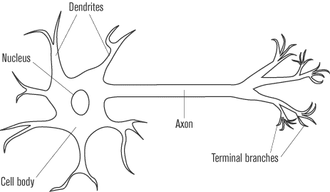 A 67: The diagram of Neron unit of human body is shown on left.
Q 68: What do you understand by stimuli?
A 68: Stimuli are plural of stimulus. Stimuli mean events or occurrences in the environment of an organism that influence its corresponding behavior.
Q 69: What can be the maximum size of an individual nerve cell?
A 69: The maximum size of an individual nerve cell may be up to a metre long.
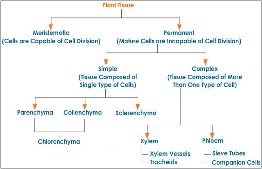
Q 70: Draw a chart to show various types of plant tissues.
A 70: The chart showing various types of plant tissues is shown: